39 lateral view of skull with labels
Molecularly defined circuits for cardiovascular and cardiopulmonary ... a, Strategy for labelling ACP or ACV cardiac terminals by delivering AAV-DIO-eYFP encoding Cre-dependent eYFP reporter (green) to left (L) or right (R) Amb in Calb1cre (ACP driver) or Ghsrcre (ACV... Normal pediatric imaging examples | Radiology Reference Article ... This article lists examples of normal imaging of the pediatric patients divided by region, modality, and age. Chest Plain radiograph chest radiograph premature (27 weeks): example 1 neonate: example 1 (lateral decubitus) 6-year-old: examp...
Thorax Abdomen and Posterior Back Muscles - Assignment Worker Activities Muscles of the Thorax, Abdomen, and Posterior Back Don't use plagiarized sources. Get Your Custom Essay on Thorax Abdomen and Posterior Back Muscles Just from $10/Page Order Essay Associated SLOs 1. Identify and define anatomical features of the spinal cord on a model or diagram for both longitudinal view and cross-sectional views. 2. Apply… Continue reading Thorax Abdomen and ...

Lateral view of skull with labels
Multi sequence average templates for aging and neurodegenerative ... This difference was most prominent in the V-AD group, for which the left and right lateral ventricles were 33% and 43% larger respectively for the male template (Table 5). Regarding asymmetry, in ... Anatomy Project - Sheridan College Neck. · Connecting the shaft and head of the femur. · Projects superior and medial from the shaft to the head. · In addition to projecting superior and medial from the shaft of the femur, the neck also projects somewhat anterior. · The amount of forward projection is extremely variable, but on an average is from 12° to 14°. Sheridan College - Dog Skeletal Anatomy Scapula. Medial View of Scapula. Clincal Aspect. The scapula is a flat triangular bone at the top of the shoulder; more commonly known as the shoulder blade. It consists of 2 surfaces (medial and lateral), 3 borders (cranial, caudal and dorsal) and 3 angles (craniodorsal, caudodorsal and ventral angle). The pig and horse do not have an acromion.
Lateral view of skull with labels. Spinal Cord Cross Section Explained (with Videos ... - New Health Advisor In the center of the gray matter you will find the cerebrospinal fluid. The specific horns of the gray matter are responsible for different things. Each horn has different functions. The dorsal and ventral horns have the neurons that direct the skeletal muscles; the lateral horn has the cells that work the smooth muscle and supply cardiac. 3. Tobacco packaging warning messages - Wikipedia Tobacco package warning messages are warning messages that appear on the packaging of cigarettes and other tobacco products concerning their health effects. They have been implemented in an effort to enhance the public's awareness of the harmful effects of smoking. In general, warnings used in different countries try to emphasize the same messages. Thoracic Disc Herniation - What You Need to Know - Drugs.com Thoracic disc herniation occurs when a thoracic disc bulges out from between your vertebrae. Discs are spongy cushions between the vertebrae in your spine. Thoracic discs are located between the base of your neck and your lower back. The herniated disc may press on your nerves or spinal cord. 10 333 part O ww Label the bones of the skull in lateral view Maxi ... 10 333 part O ww Label the bones of the skull in lateral view Maxi Frontal bone Occipital bone Temporal bone Nasal bone Pantal bone Sphenoid bone Zygomatic bone Mandible Zoom 11 3.33 points Label the specific bony features of the skull in midsagittal view Cribriform plate Vomer Sella turcica Angle of mandible Perpendicular Reset Zoom Skull M
EOF Bones Of The Upper Limb Quiz - ProProfs The upper limb is made up of a total of about sixty-four bones, and most are located in our hands. The quiz below is designed to see how well you know the bones of the upper limb. It will test out your knowledge about it, and you will also get to learn more through this quiz. Best of luck and have fun! Questions and Answers 1. Positions and Functions of the Four Brain Lobes - MD-Health.com The occipital lobe, the smallest of the four lobes of the brain, is located near the posterior region of the cerebral cortex, near the back of the skull. The occipital lobe is the primary visual processing center of the brain. Here are some other functions of the occipital lobe: Visual-spatial processing Movement and color recognition Lateral ventricle volume trajectories predict response inhibition in ... Several processing steps, such as skull 130 stripping, Talairach transforms, atlas registration as well as spherical surface maps and 131 parcellations are then initialized with common information from the within-subject 132 template, significantly increasing reliability and statistical power [44].
DePuy Synthes | EMEA | J&J MedTech DePuy Synthes solutions, in specialties including joint reconstruction, trauma, craniomaxillofacial, spinal surgery and sports medicine, are designed to advance patient care while delivering clinical and economic value to health care systems worldwide. As we celebrate 125 years of innovation, our commitment is to keep people moving. Antenatal Care Module: 3. Anatomy and Physiology of the Female ... The vestibule is the area between the labia minora, and consists of the clitoris, urethral opening and the vaginal opening. The clitoris is a short erectile organ at the top of the vestibule, which has a very rich nerve supply and blood vessels. Its function is sexual excitation and it is very sensitive to touch. 11 Possible Causes of Shooting Pain on Head - New Health Advisor Whenever you use medications, be sure that you are not taking more than the recommended dosage on the label. Other ways that you can release the tension headache is by applying a heating pad, taking a warm shower, getting some sleep, or getting a message to loosen the tense muscles. 3. Ice Pick Headaches Co-registration of mouse cortical structures between in vivo and ex ... Alignment between in vivo and ex vivo images to locate the field of view in both image volumes (A) in vivo widefield image of a 5 mm cranial window over a mouse's posterior cortical surface.
Skull diagram, lateral view with labels part 2 - Axial Ske… | Flickr
Bev Fitchett's Guns 1 With the rifle fully unloaded, and sight set at zero windage and 100 yards elevation, as explained in TM 9-720, remove the bolt from the rifle. 2 Place the rifle on a solid rest and, looking through the bore, aline the center of the bore with a point about 100 yards distant. 3 Without shifting the rifle, look through the sight and shift the rear end by means of the lateral adjustment screws ...

Transcranial View of the Skull Base | Neuroanatomy | The Neurosurgical ...
Sheridan College - Dog Skeletal Anatomy Scapula. Medial View of Scapula. Clincal Aspect. The scapula is a flat triangular bone at the top of the shoulder; more commonly known as the shoulder blade. It consists of 2 surfaces (medial and lateral), 3 borders (cranial, caudal and dorsal) and 3 angles (craniodorsal, caudodorsal and ventral angle). The pig and horse do not have an acromion.
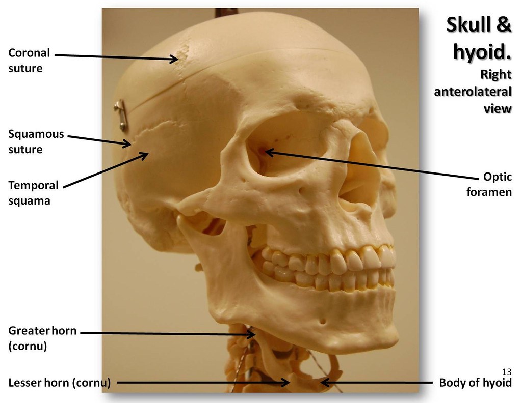
Skull, anterolateral view with labels - Axial Skeleton Vis… | Flickr
Anatomy Project - Sheridan College Neck. · Connecting the shaft and head of the femur. · Projects superior and medial from the shaft to the head. · In addition to projecting superior and medial from the shaft of the femur, the neck also projects somewhat anterior. · The amount of forward projection is extremely variable, but on an average is from 12° to 14°.

Lateral View of the Skull | Natalie Cormier
Multi sequence average templates for aging and neurodegenerative ... This difference was most prominent in the V-AD group, for which the left and right lateral ventricles were 33% and 43% larger respectively for the male template (Table 5). Regarding asymmetry, in ...

Inferior View of Skull Quiz (KNES 259)
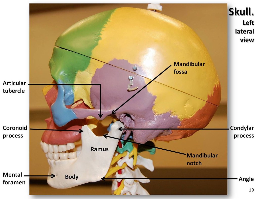
Multi-colored Skull, lateral view with labels - Axial Skel… | Flickr

MBBS Medicine (Humanity First): Skull Anatomy

Lateral View of the Brain | ClipArt ETC
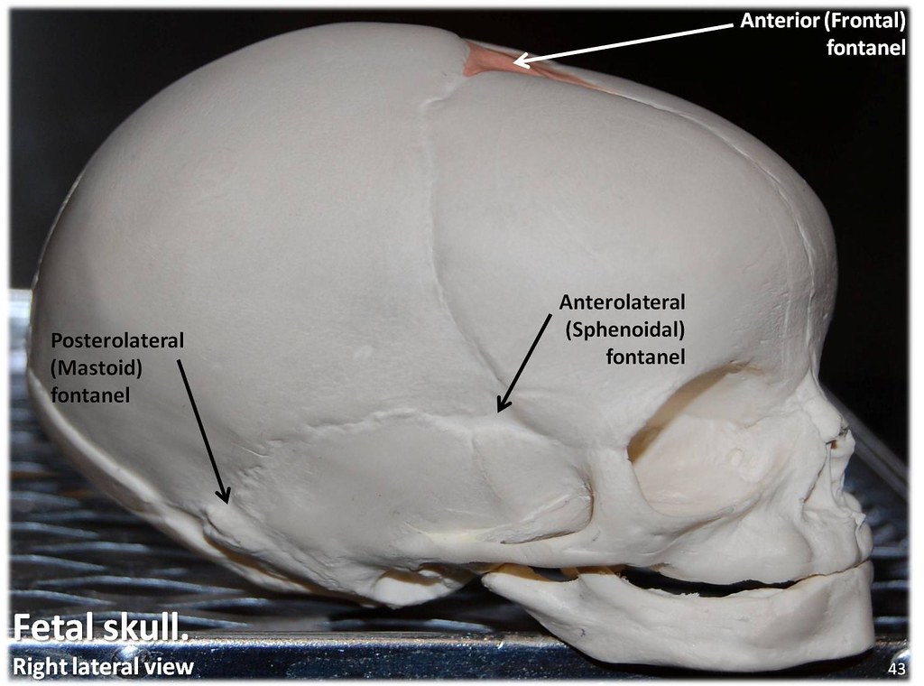
Fetal Skull, lateral view with labels - Axial Skeleton Visual Atlas ...
Post a Comment for "39 lateral view of skull with labels"