43 labels of the human brain
File:Brain human normal inferior view with labels en.svg Size of this PNG preview of this SVG file: 424 × 505 pixels. Other resolutions: 201 × 240 pixels | 403 × 480 pixels | 645 × 768 pixels | 860 × 1,024 pixels | 1,719 × 2,048 pixels. Original file (SVG file, nominally 424 × 505 pixels, file size: 169 KB) File information. Structured data. Captions. Propagating labels of the human brain based on non-rigid MR image ... Background: In order to perform statistical analysis of cohorts based on images, reliable methods for automated anatomical segmentation are required. Label propagation (LP) from manually segmented atlases onto newly acquired images is a particularly promising approach. Methods: We investigated LP on a set of 6 three-dimensional T1-weighted magnetic resonance data sets of the brains of normal ...
The Human Brain | Brain and Cognitive Sciences | MIT OpenCourseWare This course surveys the core perceptual and cognitive abilities of the human mind and asks how they are implemented in the brain. Key themes include the representations, development, and degree of functional specificity of these components of mind and brain. The course will take students straight to the cutting edge of the field, empowering ...

Labels of the human brain
Nervous System - Label the Brain - TheInspiredInstructor.com This brain part controls involuntary actions such as breathing, heartbeats, and digestion. (12) cerebrum cerebellum brain stem spinal cord. This part of the nervous system moves messages between the brain and the body. (13) frontal occipital parietal temporal. This part of the cerebrum interprets and sorts information from the senses. Brain Anatomy and How the Brain Works - Hopkins Medicine The largest lobe of the brain, located in the front of the head, the frontal lobe is involved in personality characteristics, decision-making and movement. Recognition of smell usually involves parts of the frontal lobe. The frontal lobe contains Broca's area, which is associated with speech ability. Parietal lobe. Automated labeling of the human brain: a preliminary report on the ... These rules have been greatly refined and are given for structures within each hierarchical level of the cerebrum as follows: Level 1 (Hemisphere) Exterior boundaries defined using a convex hull of cortex Medial boundaries manually outlined from atlas sections Level 2 (Lobe) Lobes Exterior boundary identical to hemispheres
Labels of the human brain. 101 Labeled Brain Images and a Consistent Human Cortical Labeling Protocol Abstract and Figures. We introduce the Mindboggle-101 dataset, the largest and most complete set of free, publicly accessible, manually labeled human brain images. To manually label the ... Automatic labeling of cortical sulci for the human fetal brain based on ... We labeled the sulci in 25 typically developing (TD) fetuses (22.9 to 29.6 GW) and validated the labeling accuracy against manually assigned sulcal labels. As the first paper on sulci labeling in the fetal brain, we systematically assessed the MTBL against other approaches typically applied to the adult population. Human Brain - Structure, Diagram, Parts Of Human Brain Following are the major parts of the human brain: Forebrain - Largest part of the brain. It is the anterior part of the brain. The forebrain parts include: Cerebrum; Hypothalamus; Thalamus; Forebrain Function: Controls the reproductive functions, body temperature, emotions, hunger and sleep. Fact: The largest among the forebrain parts is the cerebrum. It is also the largest part of all vertebrate brains. 101 labeled brain images and a consistent human cortical labeling ... We introduce the Mindboggle-101 dataset, the largest and most complete set of free, publicly accessible, manually labeled human brain images. To manually label the macroscopic anatomy in magnetic resonance images of 101 healthy participants, we created a new cortical labeling protocol that relies on robust anatomical landmarks and minimal manual edits after initialization with automated labels.
Main Parts of the Human Brain and Subdivisions of Human Brain Parts Thalamus, epithalamus, subthalamus and hypothalamus are the four sub-divisions. Here Hypothalamus is one of the parts of the human brain that initiates, coordinates, maintains and assists in the successful accomplishment of a number of visceral activities with the help of its hormonal secretions. Diagram Of Brain with their Labelings and Detailed Explanation The parietal lobe is found at the upper back of our brain. This lobe functions by controlling all our complex behaviours, including senses of vision, the sense of touch, spatial orientation and body awareness. It manages body position, movements, the perception of stimuli, orientation, handwriting and visuospatial processing. The Occipital Lobe Solved Label the structures and lobes of the human brain by - Chegg Label the structures and lobes of the human brain by clicking and dragging the labels to the correct location. <--Anterior Posterior --> Precentral gyrus Temporal lobe Parieto-occipital sulcus Parietal lobe Lateral sulcus Insula Postcentral gyrus Central sulcus Occipital lobe Frontal lobe Reset Zoom Intriguing answers: what it is about the human brain that makes us ... This led us to the conclusion that additional human brain tissue, acquired as a result of evolution, may be primarily dedicated to synergy. In turn, it is tempting to speculate that the advantages ...
Automated labeling of the human brain: a preliminary report on the ... The accuracy of the Talairach labels is dependent on the accuracy of the spatial normalization used to conform brain images to the 1988 atlas brain. For spatial normalization methods used today, it is anticipated that label conformance will be good for structures in levels 1-3. Whole Brain Segmentation : Automated Labeling of Neuroanatomical ... In contrast to existing segmentation procedures that only label a small number of tissue classes, the current method assigns one of 37 labels to each voxel, including left and right caudate, putamen, pallidum, thalamus, lateral ventricles, hippocampus, and amygdala. Label the Human Brain - 4th Grade Science Worksheet - SoD Label the Human Brain Label the Human Brain. Your brain may look like an ugly wrinkled gray sponge but it is actually the lord and master of your body. It enables your body to run smoothly and controls everything you do, asleep or wide awake. Isn't that amazing! This fun science worksheet for 4th grade helps you learn about the different parts of the human brain anatomy. 101 Labeled Brain Images and a Consistent Human Cortical Labeling ... In this article, we introduced the largest and most complete set of free, publicly available, manually labeled human brain images - 101 human cortices labeled according to a new surface-based cortical labeling protocol. These data are available 6 under a Creative Commons (attribution-non-commercial-sharealike 3.0) license (see text footnote 3). We compared the manual labels with labels generated by automated labeling methods to set benchmarks for the evaluation of automated registration ...
PICS DOT COM: Deformation of the Human Skull
Brain (Human Anatomy): Picture, Function, Parts, Conditions, and More The brain is one of the largest and most complex organs in the human body. It is made up of more than 100 billion nerves that communicate in trillions of connections called synapses. • The ...

31 Match The Label To Its Appropriate Region Of The Brainstem. - Labels Information List
101 Labeled Brain Images and a Consistent Human Cortical Labeling Protocol We introduce the Mindboggle-101 dataset, the largest and most complete set of free, publicly accessible, manually labeled human brain images. To manually label the macroscopic anatomy in magnetic resonance images of 101 healthy participants, we created a new cortical labeling protocol that relies on robust anatomical landmarks and minimal manual edits after initialization with automated labels ...
Brain Anatomy Labeled Diagram Stock Illustration 197548709 - Shutterstock
Whole brain segmentation: automated labeling of neuroanatomical ... In contrast to existing segmentation procedures that only label a small number of tissue classes, the current method assigns one of 37 labels to each voxel, including left and right caudate, putamen, pallidum, thalamus, lateral ventricles, hippocampus, and amygdala.
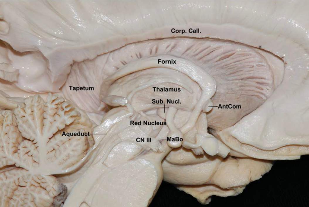
Medial View of the Left Hemisphere | Neuroanatomy | The Neurosurgical Atlas, by Aaron Cohen ...
Human brain - Wikipedia The human brain is the central organ of the human nervous system, and with the spinal cord makes up the central nervous system.The brain consists of the cerebrum, the brainstem and the cerebellum.It controls most of the activities of the body, processing, integrating, and coordinating the information it receives from the sense organs, and making decisions as to the instructions sent to the ...

30 Label Of The Brain - Labels Information List
Labeled Diagrams of the Human Brain You'll Want to Copy Now Labeled Diagrams of the Human Brain Central Core. The central core consists of the thalamus, pons, cerebellum, reticular formation and medulla. These five... Limbic System. The limbic system is present only in mammals and helps in the liaison between motivated behavior,... Cerebral Cortex. This is ...

Human Anatomy Lab: The Urinary and Reproductive Systems
Human Brain Labels Illustrations, Royalty-Free Vector Graphics & Clip ... Choose from Human Brain Labels stock illustrations from iStock. Find high-quality royalty-free vector images that you won't find anywhere else.

Human Anatomy Lab: Heart Models
Human Brain Labels Pictures, Images and Stock Photos Search from Human Brain Labels stock photos, pictures and royalty-free images from iStock. Find high-quality stock photos that you won't find anywhere else.

Diagrams - Little Medic | Labels, Human brain diagram, Brain
Automated Labeling of the Human Brain A forward-transform method for retrieving brain labels from the 1988 Talairach Atlas using x-y-z coordinates is presented. A hierarchical volume-occupancy labeling scheme was created to simplify the organization of atlas labels using volume and subvolumetric ...

Gaseous exchange in the lungs | Circulatory and respiratory systems | Siyavula
Brain: Atlas of human anatomy with MRI - e-Anatomy - IMAIOS Anatomy of the brain (MRI) - cross-sectional atlas of human anatomy. The module on the anatomy of the brain based on MRI with axial slices was redesigned, having received multiple requests from users for coronal and sagittal slices. The elaboration of this new module, its labeling of more than 524 structures on 379 MRI images in three different ...

CS379C 2018 Class Discussion Notes
Parts of the brain: Learn with diagrams and quizzes | Kenhub First up, have a look at the labeled brain structures on the image below. Try to memorize the name and location of each structure, then proceed to test yourself with the blank brain diagram provided below. Labeled diagram showing the main parts of the brain.

Label Brain Diagram | Wiring Diagram
Automated labeling of the human brain: a preliminary report on the ... These rules have been greatly refined and are given for structures within each hierarchical level of the cerebrum as follows: Level 1 (Hemisphere) Exterior boundaries defined using a convex hull of cortex Medial boundaries manually outlined from atlas sections Level 2 (Lobe) Lobes Exterior boundary identical to hemispheres

Exploration of the Human Spinal Cord
Brain Anatomy and How the Brain Works - Hopkins Medicine The largest lobe of the brain, located in the front of the head, the frontal lobe is involved in personality characteristics, decision-making and movement. Recognition of smell usually involves parts of the frontal lobe. The frontal lobe contains Broca's area, which is associated with speech ability. Parietal lobe.

Compare price to brain diagram poster | TragerLaw.biz
Nervous System - Label the Brain - TheInspiredInstructor.com This brain part controls involuntary actions such as breathing, heartbeats, and digestion. (12) cerebrum cerebellum brain stem spinal cord. This part of the nervous system moves messages between the brain and the body. (13) frontal occipital parietal temporal. This part of the cerebrum interprets and sorts information from the senses.
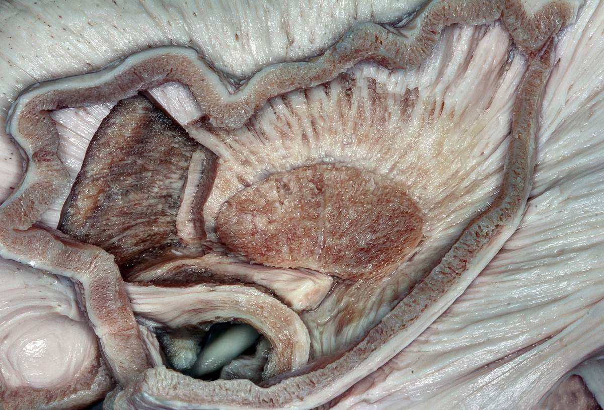
Sagittal View of the Basal Ganglia and Internal Capsule | Neuroanatomy | The Neurosurgical Atlas ...
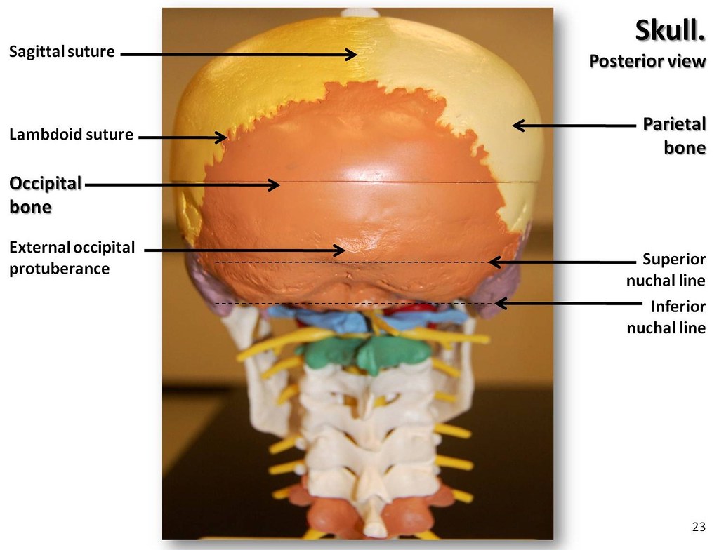
Multi-colored Skull, posterior view with labels - Axial Sk… | Flickr
Post a Comment for "43 labels of the human brain"