38 cell image with labels
How to Create 3D Plant Cell and Animal Cell Models for ... - Owlcation Take a look at some cell diagrams on an interactive site like Cells Alive. This site offers awesome animations of both plant and animal cells with descriptions of each organelle. Step 2: Choose Edible vs. Non-Edible Model Next, you should decide whether you want your cell model to be edible or not. How to Print Avery Labels from Excel (2 Simple Methods) - ExcelDemy Step 02: Make Avery Labels in Word Secondly, open a blank document in Microsoft Word. and go to the tab. Following, navigate to Mailings > Start Mail Merge > Labels. Now, choose the options as shown in the image below and click OK to close the dialog box. Next, select Design > Page Borders. Immediately, a Wizard box appears, choose Borders > Grid.
Analysis of the Human Protein Atlas Weakly Supervised Single-Cell ... The labels for each cell in the image are therefore not guaranteed to be precise because of single-cell heterogeneity. That is, in the same image of a genetically identical population, individual...

Cell image with labels
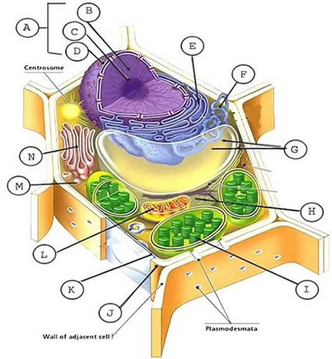
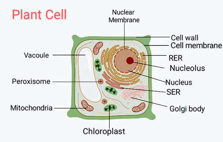
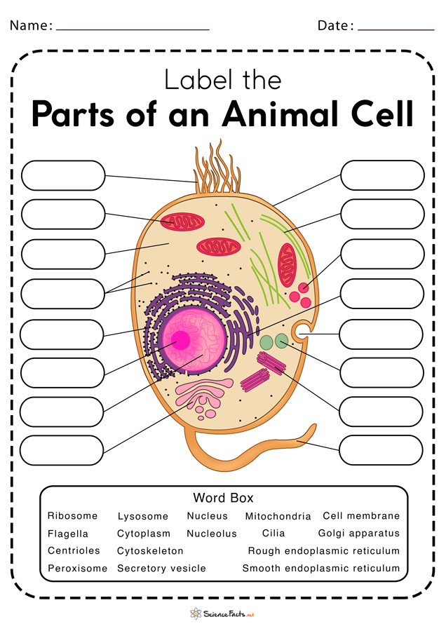
Plant Cell: Meaning, Components, Structure, Functions & Parts - Embibe These differences can be clearly understood when the cells are examined under an electron microscope. Observe the labelled diagram of plant cell structure as given below: Are Plant Cells Prokaryotic or Eukaryotic? The cell is the basic structural and functional unit of life in all living organisms. Cell Organelles- Definition, Structure, Functions, Diagram A cell wall is multilayered with a middle lamina, a primary cell wall, and a secondary cell wall. The middle lamina contains polysaccharides that provide adhesion and allow binding of the cells to one another. After the middle lamina is the primary cell wall which is composed of cellulose. Animal Cell Labeling Quiz Questions And Answers - ProProfs Take this animal cell labeling quiz to learn more about this topic! Animal cell labeling can be tricky at first, so why don't we start you off relatively easy with this animal cell part labeling quiz! In this one, we'll be giving you a question referring to a given diagram and asking you to label it. Simple, right? Let's find out how many you get right. All the best! Let's go!
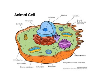
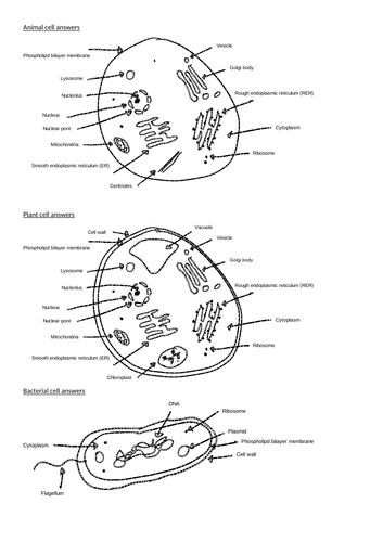
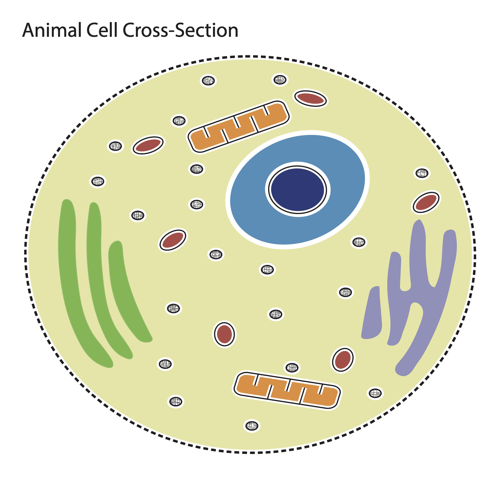
Cell image with labels. › Avery-Labels-White-Matte-Spine › dpAmazon.com : Avery CD Labels, Print to the Edge, Permanent ... These labels work great with the Avery CD Stomper. If you intend to make many CD labels, I would highly recommend getting the CD Stomper in conjunction with these labels! It really is a no brainer as long as you follow the instructions. The labels adhere to the optical disk perfectly. I have an Epson ET-16500 printer that I use to create the ... › 509290 › how-to-use-cell-valuesHow to Use Cell Values for Excel Chart Labels - How-To Geek Mar 12, 2020 · If these cell values change, then the chart labels will automatically update. Link a Chart Title to a Cell Value. In addition to the data labels, we want to link the chart title to a cell value to get something more creative and dynamic. We will begin by creating a useful chart title in a cell. We want to show the total sales in the chart title. Animal Cells: Labelled Diagram, Definitions, and Structure - Research Tweet The endoplasmic reticulum (s) are organelles that create a network of membranes that transport substances around the cell. They have phospholipid bilayers. There are two types of ER: the rough ER, and the smooth ER. The rough endoplasmic reticulum is rough because it has ribosomes (which is explained below) attached to it. ListView - .NET MAUI | Microsoft Learn ImageCell, which displays an image with primary and secondary text on separate lines. SwitchCell, which displays text and a switch that can be switched on or off. EntryCell, which displays a label and text that's editable. ViewCell, which is a custom cell whose appearance is defined by a View.
Image classification and object detection using Amazon Rekognition ... The following is a sample image from this dataset, which is included as part of the notebook. Multi-label image classification - This dataset demonstrates how to classify images into multiple categories, such as the color, size, texture, and type of a flower. For example, plant growers can use Rekognition Custom Labels to distinguish between ... Label-free prediction of cell painting from brightfield images Cell Painting is a high-content image-based assay applied in drug discovery to predict bioactivity, assess toxicity and understand mechanisms of action of chemical and genetic perturbations. We investigate label-free Cell Painting by predicting the five fluorescent Cell Painting channels from bright … › cells › bactcellInteractive Bacteria Cell Model - CELLS alive In the space are enzymes and other proteins that help digest and move nutrients into the cell. Cell Wall: Composed of peptidoglycan (polysaccharides + protein), the cell wall maintains the overall shape of a bacterial cell. The three primary shapes in bacteria are coccus (spherical), bacillus (rod-shaped) and spirillum (spiral). Self-Supervised Learning of Phenotypic Representations from Cell Images ... We propose WS-DINO as a novel framework to use weak label information in learning phenotypic representations from high-content fluorescent images of cells. Our model is based on a knowledge distillation approach with a vision transformer backbone (DINO), and we use this as a benchmark model for our study. Using WS-DINO, we fine-tuned with weak label information available in high-content ...
Interphase- Definition, Stages, Cell cycle, Diagram, Video The synthesis (S) phase is the phase of cell copying or cell duplication of its DNA of its entire genome. Gap 1 (G1) This is the phase in which the cell undergoes normal growth and cell function synthesizing high amounts of proteins. The cell increases in size and volume as more cell organelles are produced. Cell Browser Hide labels c l; Tools. Remove all custom annotations; Set as background cells b s; Reset background cells b r; Help. About; How to use this website; Interactive Tutorial; Setup your own cell browser + Learn the parts of a cell with diagrams and cell quizzes For this exercise we'll start with an image of a cell diagram ready labeled. Study this and make sure that you're clear about which structure is found where. Cell diagram unlabeled It's time to label the cell yourself! As you fill in the cell structure worksheet, remember the functions of each part of the cell that you learned in the video. Plant Cell- Definition, Structure, Parts, Functions, Labeled Diagram Functions of the plant cell (plasma) membrane. In-plant cells the cell membrane separated the cytoplasm from the cell wall. It has a selective permeability hence it regulates the contents that move in and out of the cell. It also protects the cell from external damage and provides support and stability to the cell.
Examples of different types of cells and labels. (a) shows ...
How to Insert a Picture in Microsoft Excel - How-To Geek Follow the same process as above to insert a picture in Excel. Then, click and drag a corner or edge to resize the image so that it fits within the cell where you want to place it. Alternatively, you can use the Size section in the ribbon on the Picture Format tab and use the Crop feature if necessary. You can also resize the cell if needed by ...

Labelling a cell Diagram | Quizlet
TableView - .NET MAUI | Microsoft Learn ImageCell, which displays an image with primary and secondary text on separate lines. SwitchCell, which displays text and a switch that can be switched on or off. EntryCell, which displays a label and text that's editable. ViewCell, which is a custom cell whose appearance is defined by a View.
Cells: Animal & Plant Cells With Organelle Labels by Kalin ...
How to mail merge and print labels from Excel - Ablebits.com Select document type. The Mail Merge pane will open in the right part of the screen. In the first step of the wizard, you select Labels and click Next: Starting document near the bottom. (Or you can go to the Mailings tab > Start Mail Merge group and click Start Mail Merge > Labels .) Choose the starting document.
Cell Labeling (Remote)
Top 20 Data Labeling Tools: In-depth Guide in 2022 - AIMultiple A data labeling tool is software that can find raw data in image, text, and audio formats and help data analysts label data according to specific techniques such as bounding box, landmarking, polyline, named entity recognition, etc. to prepare high-quality data for ML model training. Why are data labeling tools important?

Free Animal Cell Unlabeled, Download Free Animal Cell ...
› help › matlabDatastore for image data - MATLAB - MathWorks Image file extensions, specified as the comma-separated pair consisting of "FileExtensions" and a character vector, cell array of character vectors, string scalar, or string array. The specified extensions do not require an imformats format, and you can use the empty quotes "" to represent files without extensions.

Animal & Plant Cell: Label the Diagram and Differences Table ...
Plant and Animal Cell: Labeled Diagram, Structure, Function - Embibe Plant Cell: Plant cells are eukaryotic cells with a true nucleus along with specialized structures called organelles that carry out certain specific functions. Animal Cell: An animal cell is a type of eukaryotic cell that lacks a cell wall and has a true, membrane-bound nucleus along with other cellular organelles.

Draw a plant cell and label the parts which determine: A.The ...
Label-free prediction of cell painting from brightfield images - Nature Cell Painting is a high-content image-based assay applied in drug discovery to predict bioactivity, assess toxicity and understand mechanisms of action of chemical and genetic perturbations. We...
Cell label - Teaching resources
Blood Histology Slides with Description and Labeled Diagram Get more blood cells microscope pictures on social media of anatomy learner. Hematopoiesis - Blood cell formation. The bone marrow is the primary production site for all blood cell -lines called the pluripotential hemopoietic stem cells. The blood cell formation is a very complex process (known as hematopoiesis).
Animal and Plant Cell Labeling
Drag the correct labels to the image. Not all labels will be used ... answered • expert verified Drag the correct labels to the image. Not all labels will be used. Ricky notices a similarity between a fried egg and the diagram of a cell. Which cell organelles is Ricky likely to associate with the parts of the egg? centriole cytoplasm nucleus chloroplast plasma membrane endoplasmic reticulum

Animal Cell Labeling.pdf - cell membrane lysosome ribosome ...
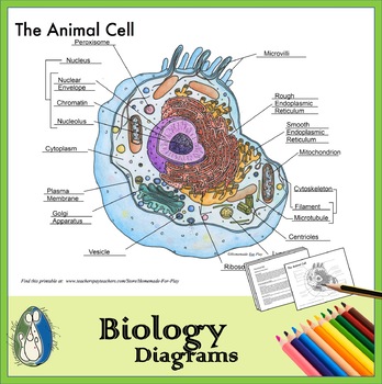
Plant Cells Vs. Animal Cells (With Diagrams) - Owlcation The most important structures of plant and animal cells are shown in the diagrams below, which provide a clear illustration of how much these cells have in common. The significant differences between plant and animal cells are also shown, and the diagrams are followed by more in-depth information. Diagram of an animal cell Doc Sonic

Cell Structure Label Organ Cell Stock Vector (Royalty Free ...
› en-us › productZE5 Cell Analyzer | Bio-Rad The ZE5 Cell Analyzer is an innovative flow cytometer with flexible configurations to meet a broad range of experimental complexities and throughput needs – accessible for novice flow cytometry users yet flexible enough for the most experienced flow cytometry professionals.

File:Plant cell structure svg labels.svg - Wikimedia Commons
Image Segmentation using Python's scikit-image module Explanation: By using rgb2gray() function, the 3-channel RGB image of shape (400, 600, 3) is converted to a single-channel monochromatic image of shape (400, 300).We will be using grayscale images for the proper implementation of thresholding functions. The average of the red, green, and blue pixel values for each pixel to get the grayscale value is a simple approach to convert a color picture ...
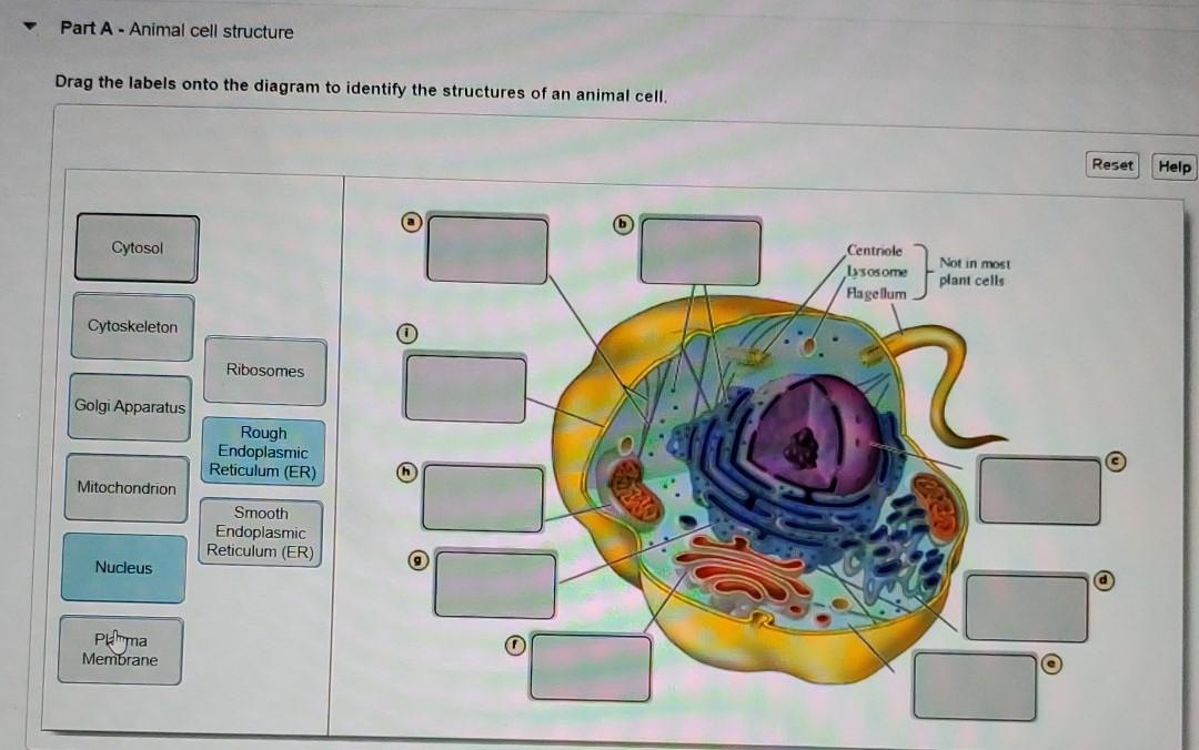
Solved Part A - Animal cell structure Drag the labels onto ...
Cells Diagram | Science Illustration Solutions - Edrawsoft Cells Diagram. Cells are the basic building blocks of all living things. The human body is composed of trillions of cells. Cells have many parts, each with a different function. Some of these parts, called organelles, are specialized structures that perform certain tasks within the cell. Drawing cells diagram helps you better understand your ...

Label the Animal Cell Worksheets (SB11866) | Animal cells ...
Using tf.keras.utils.image_dataset_from_directory with label list from the document image_dataset_from_directory it specifically required a label as inferred and none when used but the directory structures are specific to the label name. I am using the cats and dogs image to categorize where cats are labeled '0' and dog is the next label. [ Sample ]:
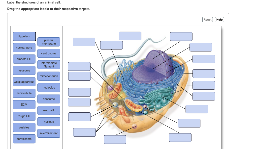
Solved Label the structures of an animal cell. Drag the ...
How to Print Labels from Excel - Lifewire Select Mailings > Write & Insert Fields > Update Labels . Once you have the Excel spreadsheet and the Word document set up, you can merge the information and print your labels. Click Finish & Merge in the Finish group on the Mailings tab. Click Edit Individual Documents to preview how your printed labels will appear. Select All > OK .
Cell Organelles Illustration Labels Stock Vector (Royalty ...
› en › productWhat's new in think-cell :: think-cell think-cell's subscription-based licensing model allows us to continuously improve our software with no additional costs for our customers. We regularly release free updates that contain great new features besides the usual improvements in stability and speed. This way think-cell becomes more powerful, more efficient and above all easier to use ...
A Vector Illustration of an Animal Cell Stock Vector ...
Simple Squamous Epithelium under a Microscope with a Labeled Diagram ... From the lung parenchyma labeled diagram, you might identify the following structures - Simple squamous epithelium lining of the lung alveoli (within the parenchyma), A connective tissue basement membrane beneath the simple squamous epithelium lining, The lumen of the lung alveoli, and The cytoplasm of the simple squamous epithelium cells.
Cell Labeling (Remote Edition) with Key
Using Custom cells to customize UITableViewCells Open the storyboard and for the CountryCell of the TableViewController, configure Custom as Cell Style: Drag two Labels and an Image View from the Library to the cell and place them like this: Select the Image View and configure the Content Mode Aspect Fill so that images are resized to fit into the image view: For both the Labels use Add New ...

Eukaryotic cells LO:to be able to label a diagram of a ...
Animal Cell Labeling Quiz Questions And Answers - ProProfs Take this animal cell labeling quiz to learn more about this topic! Animal cell labeling can be tricky at first, so why don't we start you off relatively easy with this animal cell part labeling quiz! In this one, we'll be giving you a question referring to a given diagram and asking you to label it. Simple, right? Let's find out how many you get right. All the best! Let's go!

Plant Cell and Animal Cell Diagram Quiz
Cell Organelles- Definition, Structure, Functions, Diagram A cell wall is multilayered with a middle lamina, a primary cell wall, and a secondary cell wall. The middle lamina contains polysaccharides that provide adhesion and allow binding of the cells to one another. After the middle lamina is the primary cell wall which is composed of cellulose.

IXL | Plant cell diagrams: label parts | 7th grade science
Plant Cell: Meaning, Components, Structure, Functions & Parts - Embibe These differences can be clearly understood when the cells are examined under an electron microscope. Observe the labelled diagram of plant cell structure as given below: Are Plant Cells Prokaryotic or Eukaryotic? The cell is the basic structural and functional unit of life in all living organisms.
Biology cell organelle labels A-level | Teaching Resources
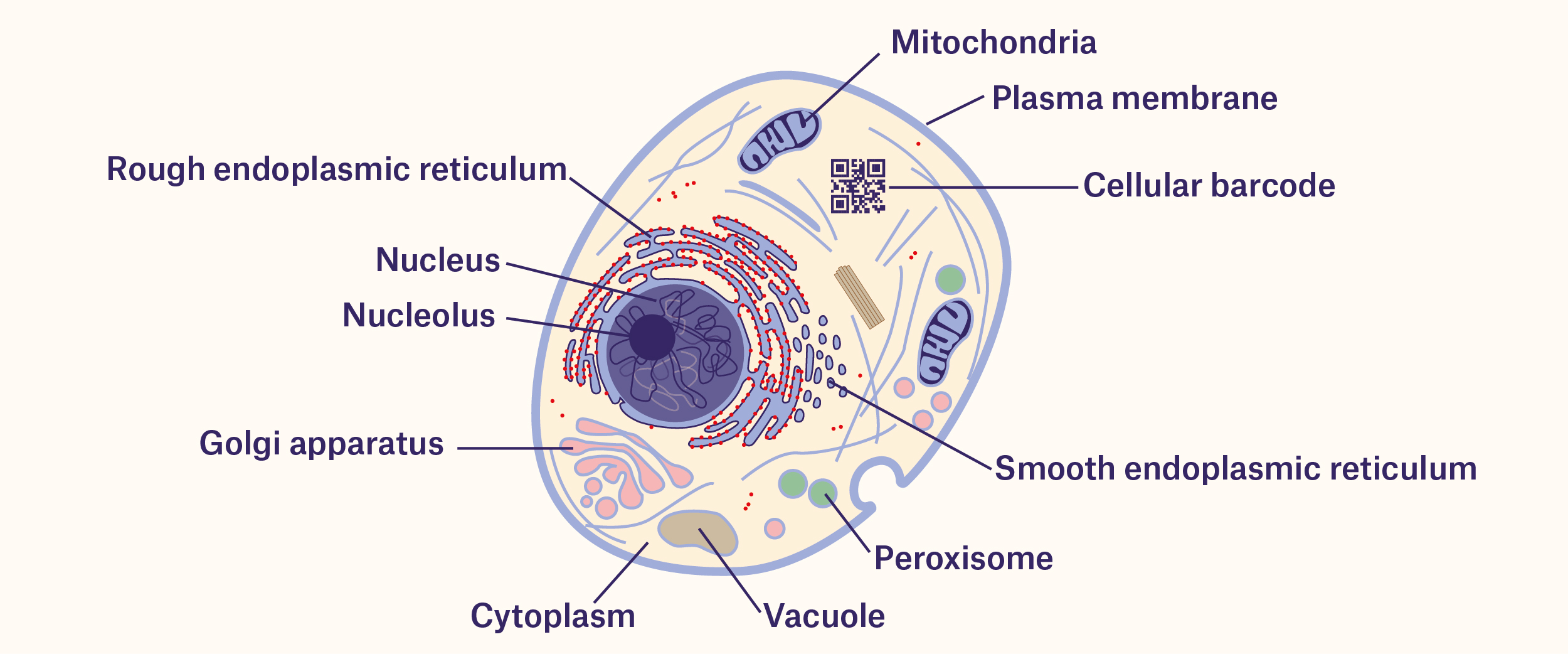
Tracking Cells With Barcodes: Beyond the Label - Labtag Blog
Plant cell label - Labelled diagram
Animal Cell Diagrams for Coloring and Labeling, with ...

Labeling of Animal cell worksheet
Free Animal Cell Unlabeled, Download Free Animal Cell ...

Animal Cell (Labels and Definition) Diagram | Quizlet

Animal Cell Labeling
Draw a diagram of a plant cell and label at least eight class ...
Animal Cell - resource - Imageshare

Cell Organelles Labeling worksheet
Animal Cell Worksheets - Free Printable
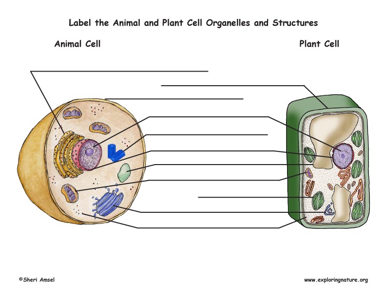
Plant and Animal Cell Labeling (Color)

Jennifer's Human Cell Label.

Dual-Activatable Cell Tracker for Controlled and Prolonged ...
Cell Labeling Page

Label-free imaging of live cells | CytoSMART
Post a Comment for "38 cell image with labels"