42 light microscope with labels
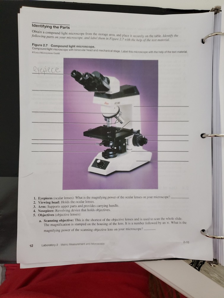
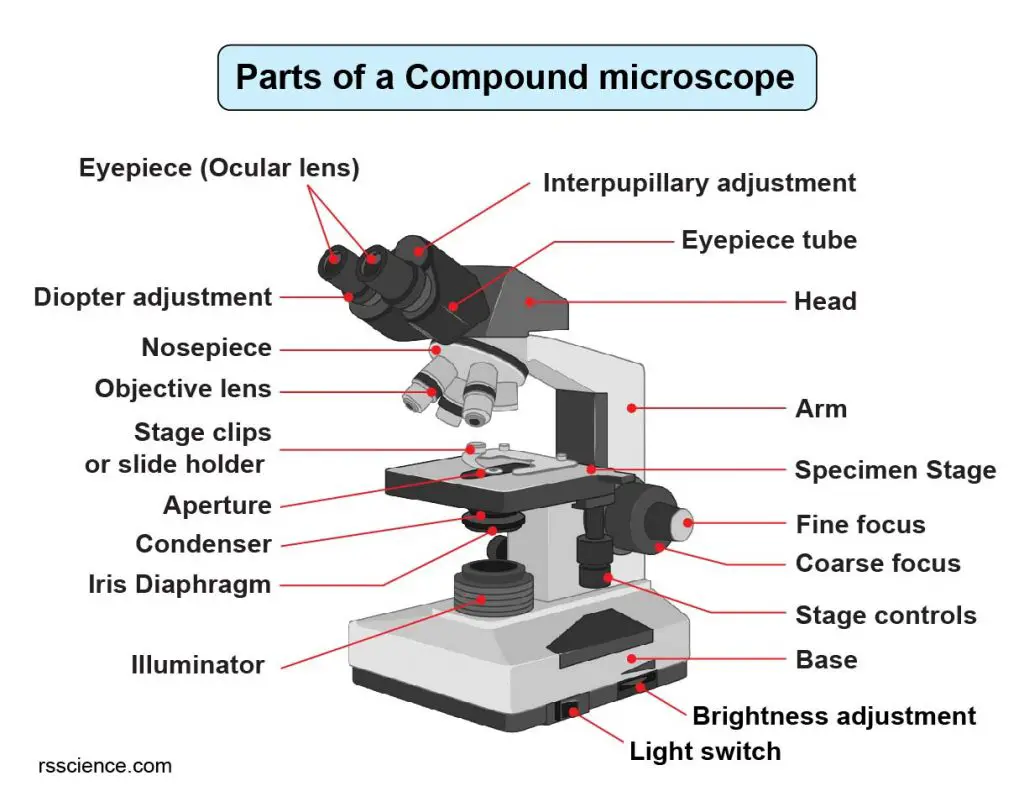
Microscope Labeling - The Biology Corner 1) Start with scanning (the shortest objective) and only use the COARSE knob . Once it is focused… 2) Switch to low power (medium) and only use the COARSE knob . You may need to recenter your slide. Once it is focused.. 3) Switch to high power (long objective). Compound Microscope Parts, Functions, and Labeled Diagram Compound Microscope Definitions for Labels Eyepiece (ocular lens) with or without Pointer: The part that is looked through at the top of the compound microscope. Eyepieces typically have a magnification between 5x & 30x. Monocular or Binocular Head: Structural support that holds & connects the eyepieces to the objective lenses.
Fluorescence Microscopy - Explanation and Labelled Images A fluorescence microscope is used to study organic and inorganic samples. Fluorescence microscopy uses fluorescence and phosphorescence to examine the structural organization, spatial distribution of samples. It is particularly used to study samples that are complex and cannot be examined under conventional transmitted-light microscope.

Light microscope with labels
Compound Microscope Parts - Labeled Diagram and their Functions The eyepiece (or ocular lens) is the lens part at the top of a microscope that the viewer looks through. The standard eyepiece has a magnification of 10x. You may exchange with an optional eyepiece ranging from 5x - 30x. [In this figure] The structure inside an eyepiece. The current design of the eyepiece is no longer a single convex lens. › microscopy › enZEISS Lightsheet 7 – Light Sheet Microscope Which optical clearing method you choose will depend on the tissue, your fluorescent labels, and the size of the sample. Lightsheet 7 is designed to match all these conditions. Image specimens at up to 2 cm in size at any refractive index between 1.33 and 1.58, and in almost all clearing solutions. Light Microscope- Definition, Principle, Types, Parts, Labeled Diagram ... A light microscope is a biology laboratory instrument or tool, that uses visible light to detect and magnify very small objects and enlarge them. They use lenses to focus light on the specimen, magnifying it thus producing an image. The specimen is normally placed close to the microscopic lens.
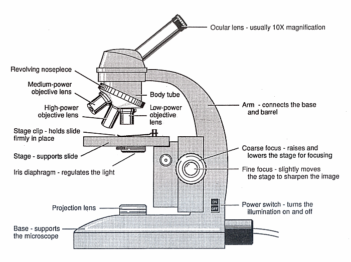
Light microscope with labels. Parts of a microscope with functions and labeled diagram - Microbe Notes Microscopic illuminator - This is the microscopes light source, located at the base. It is used instead of a mirror. It captures light from an external source of a low voltage of about 100v. Condenser - These are lenses that are used to collect and focus light from the illuminator into the specimen. A Study of the Microscope and its Functions With a Labeled Diagram ... To better understand the structure and function of a microscope, we need to take a look at the labeled microscope diagrams of the compound and electron microscope. These diagrams clearly explain the functioning of the microscopes along with their respective parts. Man's curiosity has led to great inventions. The microscope is one of them. Microscope Labeled Pictures, Images and Stock Photos Diagram of the process of photosynthesis, showing the light reactions and the Calvin cycle. photosynthesis by absorbing water, light from the sun, and carbon dioxide from the atmosphere and converting it to sugars and oxygen. Light reactions occur in the thylakoid. Calvin Cycle occurs in the stoma. microscope labeled stock illustrations Binocular Microscope Anatomy - Parts and Functions with a Labeled ... Ocular lens or eyepiece of the microscope, Diopter adjustment of the eyepiece All of these parts are identified in a light microscope labeled diagram. So, first, make sure you can identify all these parts from this labeled diagram. Parts of the compound microscope
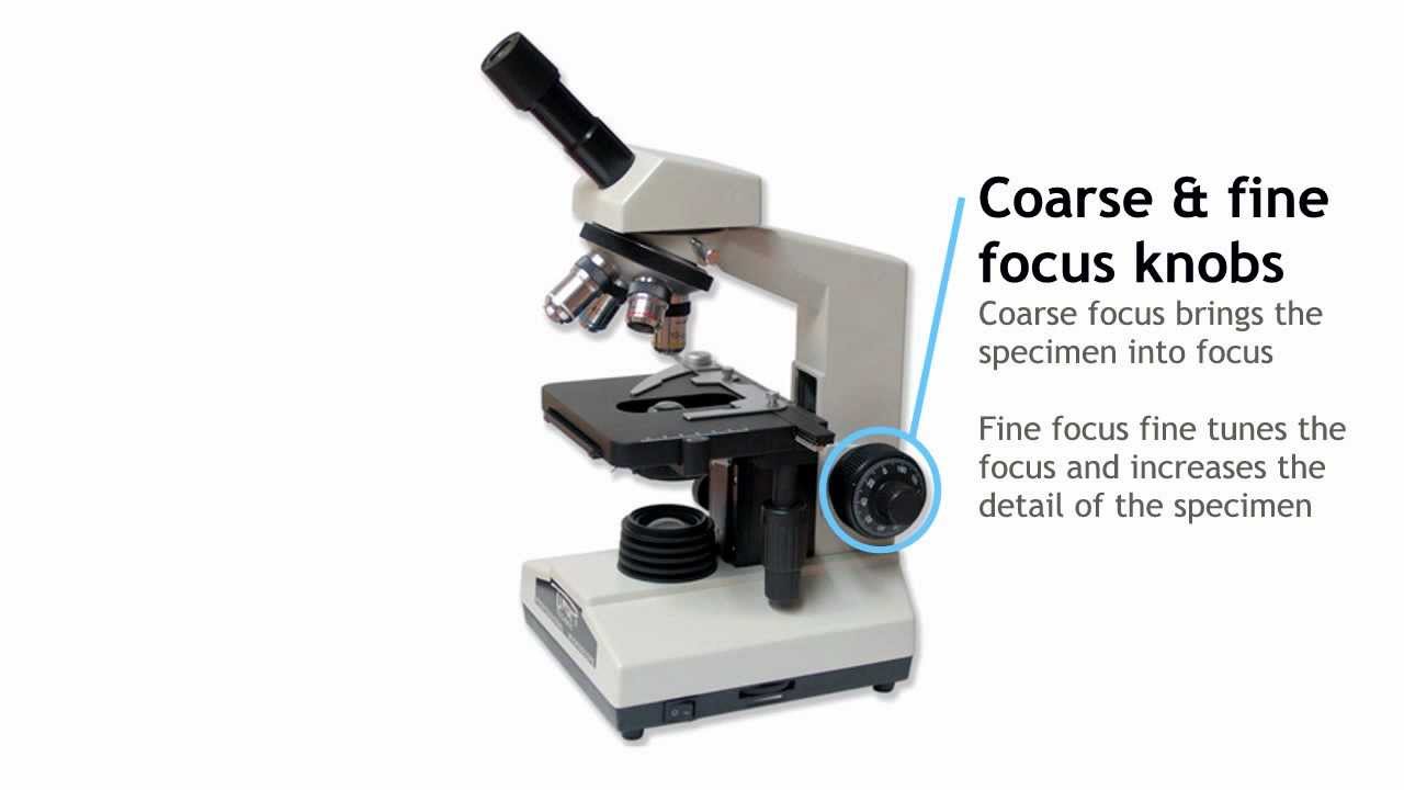
Label the Light Microscope - Labelled diagram - Wordwall Drag and drop the pins to their correct place on the image.. Eyepiece, Light Source, Base, Stage, Stage Clips, Fine Focus, Coarse Focus, Arm, Objective Lens. Parts of the Microscope with Labeling (also Free Printouts) Home Microscopes Parts of the Microscope with Labeling (also Free Printouts) By Editorial Team March 7, 2022 A microscope is one of the invaluable tools in the laboratory setting. It is used to observe things that cannot be seen by the naked eye. Table of Contents 1. Eyepiece 2. Body tube/Head 3. Turret/Nose piece 4. Objective lenses 5. Labeling the Parts of the Microscope | Microscope World Resources Labeling the Parts of the Microscope This activity has been designed for use in homes and schools. Each microscope layout (both blank and the version with answers) are available as PDF downloads. You can view a more in-depth review of each part of the microscope here. Download the Label the Parts of the Microscope PDF printable version here. Compound Microscope Labeled Diagram | Quizlet Part that supports the microscope. Stage. Supports the slide or specimen. Coarse adjustment Knob. sed to focus when using the low power objective lenses. Fine Adjustment Knob. Used to focus the image on high power to view image in more detail. Revolving nose piece. The revolving piece on which the lenses are attached.
Light Microscope: Functions, Parts and How to Use It To use a light microscope, you can follow the steps below carefully. Start with a low lens and a clean slide. The microscope stage should be lowered as low as possible. Center the slide so that the specimen is under the objective lens. Use the coarse adjustment knob to get a general focus. Then slowly move up the stage until focus is achieved. Parts of a Microscope Labeling Activity - Storyboard That In this activity, students will create a poster of a microscope with labeled parts. Students will identify and describe the microscope parts and functions. This is an awesome activity to complete at the beginning of either the school year or the unit on basic cells. ... Provides light to illuminate the specimen, sometimes a mirror is also used ... Solved Label the image of a compound light microscope using - Chegg Expert Answer. 100% (17 ratings) Transcribed image text: Label the image of a compound light microscope using the terms provided. › NATIONAL-GEOGRAPHIC-Dual-StudentNational Geographic Dual LED Student Microscope Aug 07, 2017 · Buy NATIONAL GEOGRAPHIC Dual LED Student Microscope - 50+ pc Science Kit with 10 Prepared Biological & 10 Blank Slides, Lab Shrimp Experiment, Perfect for School Laboratory, Homeschool & Home Education: Microscopes - Amazon.com FREE DELIVERY possible on eligible purchases
Microscopy and Its Types - BIOLOGY EASE
› microscopy › enZEISS Axio Observer for Life Science Research Fast switchable light sources and filters give you highest spectral flexibility and speed. Select the ideal camera to always get the image quality and speed your applications require. Whether keeping your sample in focus for long-term imaging or adapting your objective to your sample, it's all automatic with this highly organized system.

Compound Microscope Parts – Labeled Diagram and their ...
Labelled Diagram Of A Light Microscope - GlobalSpec Products/Services for Labelled Diagram Of A Light Microscope Microscopes - (706 companies) ...and transmission electron microscopes. Acoustic and ultrasonic microscopes use sound waves to create images of the sample. Compound microscopes use a single light path. These types of microscopes can have a single eyepiece (monocular) or a dual eyepiece...

Monday 10/19/15 AIM: how do the parts of the compound light ...
› microscopy › enZEISS LSM 900 with Airyscan 2 - Compact Confocal Microscope ... Airyscan 2 is an area detector with 32 circularly arranged detection elements. Each of these acts as a small pinhole, contributing to super-resolution information, while the complete detector area collects more light than the standard confocal setting. This produces much greater light efficiency while capturing enhanced structural information.

Below is a photo of a compound light microscope with labels ...
Sperm Under Microscope with Labeled Diagram - AnatomyLearner Under the light microscope, the sperm consists of two main portions - the head and the tail. But, the electron microscope shows four different parts in the tail of spermatozoa. ... So, this article provides the details structural features of sperm under the light microscope. All the labeled diagrams might help you identify the sperms from ...
Microscopes | Idaho State University
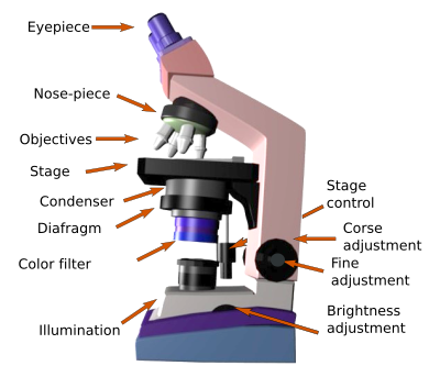
Microscope Parts, Function, & Labeled Diagram - slidingmotion Diaphragm. The diaphragm is also called as iris. This iris situates below the stage of the microscope. The function of the diaphragm is to control the amount of light that focuses on the specimen. This diaphragm can adjust the amount of light and intensity of light that falls on the specimen. In some standard and high-quality microscopes, this ...

Label a Microscope Worksheet
Compound Light Microscope: Everything You Need to Know A compound light microscope is a type of light microscope that uses a compound lens system, meaning, it operates through two sets of lenses to magnify the image of a specimen. It's an upright microscope that produces a two-dimensional image and has a higher magnification than a stereoscopic microscope.

Microscope diagram labeled | Clipart Panda - Free Clipart Images
Microscope Parts and Functions This allows the slide to be easily inserted or removed from the microscope. It also allows the specimen to be labeled, transported, and stored without damage. Stage: The flat platform where the slide is placed. Stage clips: Metal clips that hold the slide in place.
1.2: Microscopes - Biology LibreTexts
Light Microscope- Definition, Principle, Types, Parts, Labeled Diagram ... A light microscope is a biology laboratory instrument or tool, that uses visible light to detect and magnify very small objects and enlarge them. They use lenses to focus light on the specimen, magnifying it thus producing an image. The specimen is normally placed close to the microscopic lens.

Label the microscope — Science Learning Hub
› microscopy › enZEISS Lightsheet 7 – Light Sheet Microscope Which optical clearing method you choose will depend on the tissue, your fluorescent labels, and the size of the sample. Lightsheet 7 is designed to match all these conditions. Image specimens at up to 2 cm in size at any refractive index between 1.33 and 1.58, and in almost all clearing solutions.
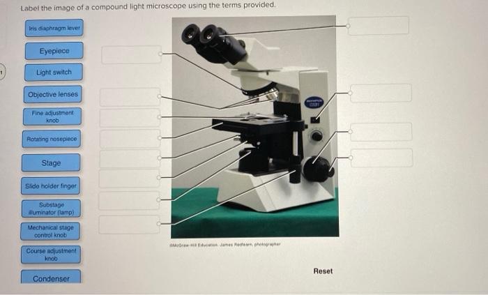
Solved Label the image of a compound light microscope using ...
Compound Microscope Parts - Labeled Diagram and their Functions The eyepiece (or ocular lens) is the lens part at the top of a microscope that the viewer looks through. The standard eyepiece has a magnification of 10x. You may exchange with an optional eyepiece ranging from 5x - 30x. [In this figure] The structure inside an eyepiece. The current design of the eyepiece is no longer a single convex lens.
Histological techniques. 6. Visualization. Light microscope ...

The Microscope Types of Microscopes Compound light microscope ...
simple light microscope labeled - Clip Art Library

Simple Microscope - Diagram (Parts labelled), Principle ...

Microscope With Labels clip art | Microscope parts ...

Microscope Labeling Activity - SMART Board Activity - Interactive Review

The Science Break - Labels for the light microscope for GCSE ...

Microscope Diagram and Quiz | Science printables, Science ...
Solved biology 1000 lab 2.7 compound light microscope ONLY ...

Microscope labeled diagram
Guide to using a microscope - Home
Microscopes: Labelling of light microscopes and difference ...
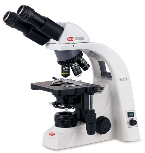
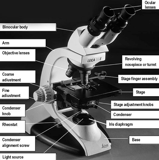
Motic® BA310E Biological Light Microscope

Microscope With Labels Clip Art at Clker.com - vector clip ...
Parts of a Compound Light Microscope

Labeling the Parts of the Microscope | Microscope activity ...

Compound Microscope Parts, Functions, and Labeled Diagram ...

Microscope Labeling #1 Diagram | Quizlet

Compound Light Microscope Labeling Diagram | Quizlet

Microscope labeled diagram
Parts of a Microscope Quiz

Living Environment Course
Label a microscope - Teaching resources
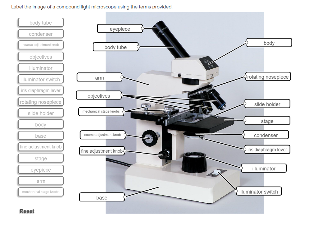
Solved Label the image of a compound light microscope using ...
Parts of a microscope with functions and labeled diagram

Label the numbered parts of the microscope - ppt download

Parts of a Microscope Labeling Activity

What is a Compound Microscope? | Microscope World Blog

Compound Microscope Parts – Labeled Diagram and their ...
What is a compound light microscope? - Dr. Biology Questions ...

Compound Microscope Parts – Labeled Diagram and their ...

label microscope diagram | Charts | Microscope, Anatomy bones ...
Post a Comment for "42 light microscope with labels"