38 human eye diagram without labels
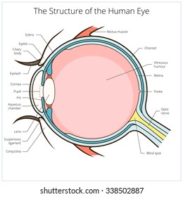
Eye Diagram With Labels and detailed description - BYJUS A brief description of the eye along with a well-labelled diagram is given below for reference. Well-Labelled Diagram of Eye The anterior chamber of the eye is the space between the cornea and the iris and is filled with a lubricating fluid, aqueous humour. The vascular layer of the eye, known as the choroid contains the connective tissue. Anatomy of the eye: Quizzes and diagrams | Kenhub Take a look at the diagram of the eyeball above. Here you can see all of the main structures in this area. Spend some time reviewing the name and location of each one, then try to label the eye yourself - without peeking! - using the eye diagram (blank) below. Unlabeled diagram of the eye
eyeball diagram to label; Eye Cross Section Labeled Diagram Stock Vector - Illustration of we have 8 Images about Eye Cross Section Labeled Diagram Stock Vector - Illustration of like Medical Stock Art, Anatomy of the Eye, Human Eye Diagram, How The Eye Work -15 Amazing Facts of Eye and also Human Eye Diagram, How The Eye Work -15 Amazing Facts of Eye. Read more:

Human eye diagram without labels
Activity Sheet 1: How the Eyes Work | Human eye diagram, Teaching ... Description Use these simple eye diagrams to help students learn about the human eye. Three differentiated worksheets are included: 1. Write the words using a word bank 2. Cut and paste the words 3. Write the words without a word bank Labels include: eyebrow, eyelid, eyelashes, pupil, iris, and sclera. Eye Test: 3 Free Eye Charts to Download and Print at Home Eye doctors can use different eye test charts for different patients and situations. The three most common eye charts are: Snellen eye chart. "Tumbling E" eye chart. Jaeger eye chart. We've included a link to download your very own eye chart after each section below. You can print these charts and test your vision right in your own home. Eye, External Front View - resource - Imageshare - Benetech Diagram of the external view of a human eye. Design modalities for the image include braille with and without labels, print with and without labels in greyscale, color, and texture. (Source: Benetech) Metadata. Subject: Life Sciences - Science. Keywords: anatomy, diagram ...
Human eye diagram without labels. Eye Anatomy: Parts of the Eye and How We See Behind the anterior chamber is the eye's iris (the colored part of the eye) and the dark hole in the middle called the pupil. Muscles in the iris dilate (widen) or constrict (narrow) the pupil to control the amount of light reaching the back of the eye. Directly behind the pupil sits the lens. The lens focuses light toward the back of the eye. Eye Diagram Unlabelled - schematron.org Download and use them in your website, document or presentation. Best Human eye diagram unlabelled free vector download for commercial use in ai, eps, cdr, svg vector illustration graphic art design format. human eye. Ask A Biologistcoloring page | Web address:schematron.org coloring. Human Eye. Page 2. 5. 3. 2. 4. File:Diagram of human eye without labels.svg - Wikimedia Commons File:Diagram of human eye without labels.svg. Size of this PNG preview of this SVG file: 410 × 430 pixels. Other resolutions: 229 × 240 pixels | 458 × 480 pixels | 732 × 768 pixels | 976 × 1,024 pixels | 1,953 × 2,048 pixels. eye diagram with labelling Lacrimal Gland diagram - The Eye Si (gh)t we have 9 Pictures about Lacrimal Gland diagram - The Eye Si (gh)t like Internal Parts and Functions of the Eye | hubpages, Unlabeled Eye Diagram - ClipArt Best and also Unlabeled Eye Diagram - ClipArt Best. Here you go: Lacrimal Gland Diagram - The Eye Si (gh)t eyemakeart.wordpress.com lacrimal gland
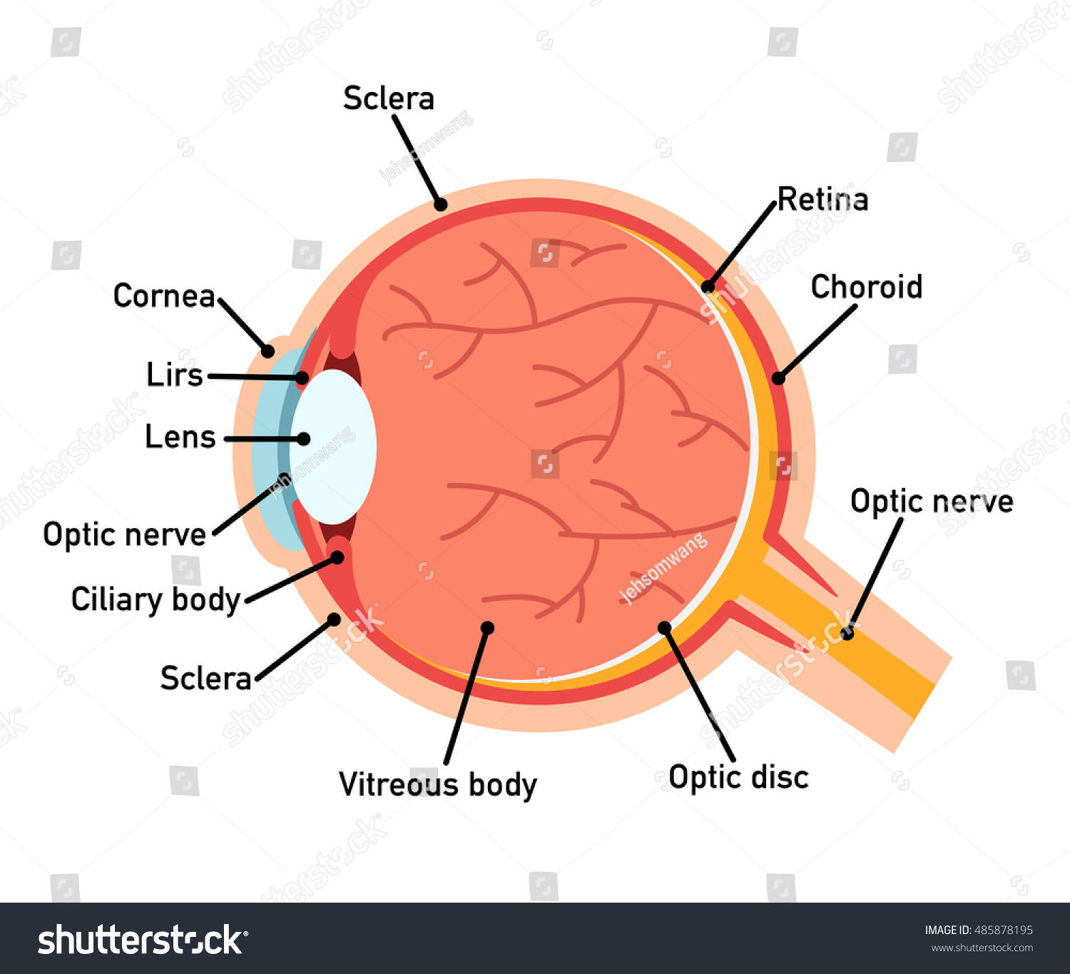
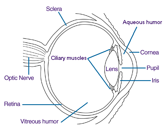
File:Schematic diagram of the human eye no.svg - Wikipedia Original upload log []. This image is a derivative work of the following images: File:Schematic diagram of the human eye en.svg licensed with PD-self 2008-02-02T01:33:45Z Jakov 508x516 (54267 Bytes) suspensory ligament, arrow was wrong; 2008-01-31T16:48:11Z Jakov 508x516 (54263 Bytes) xml-Cleanup; 2007-01-25T03:10:10Z Rhcastilhos 508x516 (42056 Bytes) {{Information |Description=Schematic ... The Eyes (Human Anatomy): Diagram, Optic Nerve, Iris, Cornea ... - WebMD The front part (what you see in the mirror) includes: Iris: the colored part. Cornea: a clear dome over the iris. Pupil: the black circular opening in the iris that lets light in. Sclera: the ... Basic Eye Anatomy - Cataract Surgery Information The following page explains basic anatomy of the human eye and highlights some structures in particular and how they relate to cataracts and cataract surgery. Figure 1.1: Normal Eye Anatomy. Eyelids and Lashes. The eyelids intermittently cover the front surface of the eye, forming a protective barrier. Blank ear diagrams and quizzes: The fastest way to learn - Kenhub Take a moment to look at the ear model labeled above. This shows you all of the structures you've just learned about in the video, labeled on one diagram. Seeing them all together in this way is a great way to learn, since anatomical structures do not exist in isolation. That's why labeling the ear is an effective way to begin your revision.
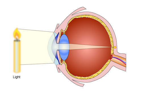
Eye anatomy: A closer look at the parts of the eye In a number of ways, the human eye works much like a digital camera: Light is focused primarily by the cornea — the clear front surface of the eye, which acts like a camera lens. The iris of the eye functions like the diaphragm of a camera, controlling the amount of light reaching the back of the eye by automatically adjusting the size of the ... Human Ear Diagram - Bodytomy Look no further, this Bodytomy article gives you a labeled human ear diagram and also explains the functions of its different components. The human body is like a big machine, and various processes take place inside it. With the help of the various organs and tissues, it carries out some of the most marvelous tasks, that are no less than a miracle! › resource › cfe2-s-12-the-humanFREE! - The Human Eye Labelling Activity - Twinkl In this resource, you’ll find a 2-page PDF that is easy to download, print out, and use immediately with your class. The first page is a labelling exercise with two diagrams of the human eye. One is a view from the outside, and the other is a more detailed cross-section. Challenge learners to label the parts of the eye diagram. On the second page, you’ll find a set of answers showing ... Eye Diagram Teaching Resources | Teachers Pay Teachers The Human Eye Overview Reading Comprehension and Diagram Worksheet. by. Teaching to the Middle. 63. $1.50. Zip. This passage briefly describes the human eye (900-1000 Lexile). 14 questions (matching and multiple choice) assess students' understanding. Students label a diagram of 6 parts of the eye. I've included a color and BW version, as well ...
Human Eyeball Diagram - YouTube
PDF Eye Anatomy Handout - National Eye Institute of light entering the eye. Lens: The lens is a clear part of the eye behind the iris that helps to focus light, or an image, on the retina. Macula: The macula is the small, sensitive area of the retina that gives central vision. It is located in the center of the retina. Optic nerve: The optic nerve is the largest sensory nerve of the eye.
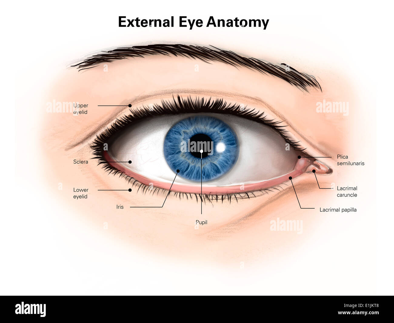
External anatomy of the human eye (with labels Stock Photo: 69866840 - Alamy
Label Parts of the Human Eye - University of Dayton Select the correct label for each part of the eye. The image is taken from above the left eye. Click on the Score button to see how you did. Incorrect answers will be marked in red.

Human Eye Anatomy Diagram Medical Description Stock Photo (Edit Now) 231982696 - Shutterstock
› 2071/1050/14-11 › 6841Human–System Interaction Based on Eye Tracking for a Virtual ... Jun 03, 2022 · With the constant exploration and development of intelligent manufacturing, the concept of digital twins has been proposed and applied. In view of the complexity and intellectualization of virtual workshop systems, real workshops can link with virtual workshosp based on AR under the structure of digital twins, which allows users to interact with virtual information and perceive the virtual ...

Human Eye Diagram Labeled - Health, Medicine and Anatomy Reference Pictures | School | Pinterest ...
Eye Diagram - Differentiated Worksheets and EASEL Activities - Pinterest Eye Diagram - Differentiated Worksheets and EASEL Activities Description Use these simple eye diagrams to help students learn about the human eye. Three differentiated worksheets are included: 1. Write the words using a word bank 2. Cut and paste the words 3.

Eye - Wikimedia Commons
BYJUS BYJUS
14 Best Images of Eye Diagram Worksheet - Human Eye Diagram Unlabeled, Anatomy and Physiology ...
anatomy human eye Anatomy eye labeled human body sclera physiology models spinal google optic nerve notes ik 4d nerves bing. ... eye diagram unlabeled unlabelled anatomy human 11e3 medical. Extreme Close-Ups Of The Human Eye - Neatorama ... Egg Emerging From An Ovary - Weird Picture Archive . hand human inside egg tendons skin without ...

Why Eating Well Isn't Enough: Part 1 - Digestion | NOURISHED WISELY
en.wikipedia.org › wiki › DopamineDopamine - Wikipedia An additional group of dopamine-secreting neurons is found in the retina of the eye. These neurons are amacrine cells, meaning that they have no axons. They release dopamine into the extracellular medium, and are specifically active during daylight hours, becoming silent at night.
Eye Anatomy Diagram,Vector Illustration. - 485878195 : Shutterstock
Eye Anatomy: Parts of the Human Eye - Vision Center The macula lutea is a yellow oval area in the retina's center (back of the eye). The center of the macula is known as the fovea. It is the section of the retina that is in charge of sharp, detailed central vision (also called visual acuity). The macula lutea has a high concentration of cones.

eye diagram
› heart › picture-of-the-heartHuman Heart (Anatomy): Diagram, Function, Chambers, Location ... Heart Tests. Electrocardiogram (ECG or EKG): A tracing of the heart’s electrical activity. Electrocardiograms can help diagnose many heart conditions. Echocardiogram: An ultrasound of the heart ...

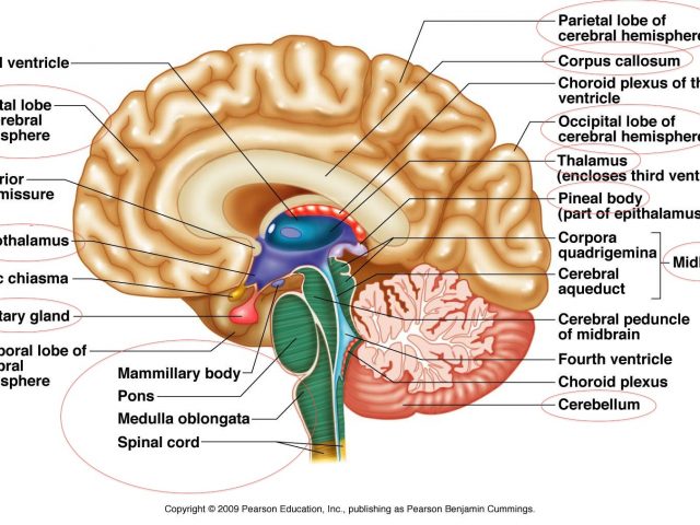
Group 4 Project: Biology: Investigating the Human eye
The Eye Diagram: What is it and why is it used? Here, the bit sequences 011, 001, 100, and 110 are superimposed over one another to obtain the example eye diagram. The eye diagram takes its name from the fact that it has the appearance of a human eye. It is created simply by superimposing successive waveforms to form a composite image. The eye diagram is used primarily to look at digital ...
Eye_Human
eyewiki.aao.org › Lens_Material_PropertiesLens Material Properties - EyeWiki The human eye does not perceive ultraviolet radiation, however the effects of ultraviolet exposure can be harmful to the eye. Transmission of ultraviolet radiation through ophthalmic lenses may be reduced or blocked depending on the lens material.
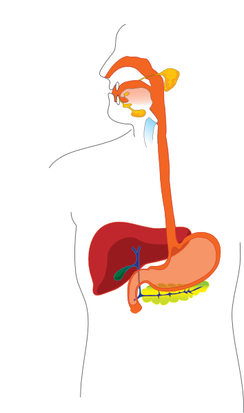
Transparent Digestive System Without Labels ~ news word
PDF Parts of the Eye - National Eye Institute | National Eye Institute Eye Diagram Handout Author: National Eye Health Education Program of the National Eye Institute, National Institutes of Health Subject: Handout illustrating parts of the eye Keywords: parts of the eye, eye diagram, vitreous gel, iris, cornea, pupil, lens, optic nerve, macula, retina Created Date: 12/16/2011 12:39:09 PM
Use of lenses for correcting vision - Pass My Exams: Easy exam revision notes for GSCE Physics
Blank Eye Diagram - Healthiack Best viewed on 1280 x 768 px resolution in any modern browser. Blank eye diagram 1063. Blank eye diagram 1020. Blank eye diagram 1023. Blank eye diagram 1029. Blank eye diagram 1031. Blank eye diagram 1033. Blank eye diagram 1034. Blank eye diagram 1035.

File:Schematic diagram of the human eye.svg - Wikimedia Commons
Ear Diagram Unlabeled - Wiring Diagrams Best Unlabeled diagram human ear free vector download for commercial use in ai, eps, cdr, svg vector illustration graphic art design format. unlabeled. Test students' knowledge of the human eye and ear as they color and label these diagrams.
Easy Human Eye Diagram With Labels - Diagram Media
en.wikipedia.org › wiki › Human_eyeHuman eye - Wikipedia Schematic diagram of the human eye. It shows a horizontal section through the right eye. The eye is made up of three coats, or layers, enclosing various anatomical structures. The outermost layer, known as the fibrous tunic, is composed of the cornea and sclera, which provide shape to the eye and support the deeper structures.
Spine Diagram With Labels - Human Anatomy
Eye, External Front View - resource - Imageshare - Benetech Diagram of the external view of a human eye. Design modalities for the image include braille with and without labels, print with and without labels in greyscale, color, and texture. (Source: Benetech) Metadata. Subject: Life Sciences - Science. Keywords: anatomy, diagram ...
Post a Comment for "38 human eye diagram without labels"